What is this project about?
This project connects mechanical characterization with biological cell culture by combining biomaterial-based 3D liver models with innovative analytical approaches. It focuses on understanding the interplay between mechanical properties and biological function in small-scale tissue constructs to improve the reliability of in vitro systems.
Your role
You will work on the development of analytical methodologies for the rheological characterization of tissue modals and cell cultures, particularly under challenging small-sample conditions.
Key tasks include
- Thin-Film Micro-Rheology:
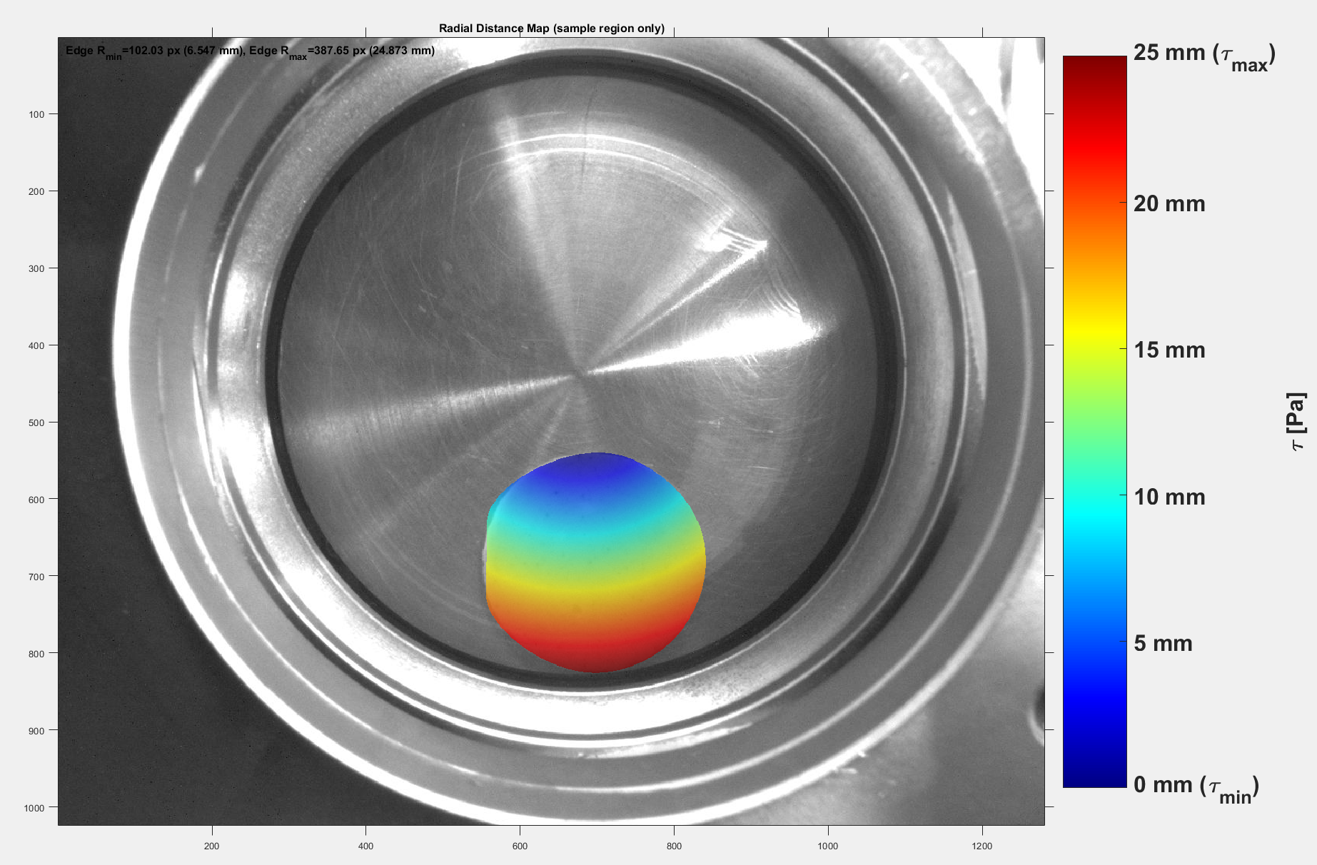
Development and implementation of rheological methods for irregular, small-scale samples (e.g., scaffolds, tissues, cell cultures), considering non-uniform sample geometries. - Optical Detection:
Integration of optical techniques to determine the contour and position of samples in situ for accurate geometry assessment during measurements. - Modeling:
Application of analytical or statistical models to calculate stress and strain distributions and improve data accuracy. - Assay Management:
Development and validation of standardized assays for reproducible measurement of biological parameters such as enzyme activity and toxicity. - Validation & Optimization:
Experimental testing, method refinement, and analysis of sample behavior under rheological conditions. - Sample Preparation:
Establishment of standardized protocols for scaffold-based cell cultures to ensure reproducibility.
Why is this important?
Conventional rheological methods are often unsuitable for small and irregular biological samples. By combining rheology with optical detection, this project aims to significantly improve the accuracy of viscoelastic measurements and contribute to more reliable in vitro models.
Focus Areas
Biomechanics · Biology · Rheology · Process Optimization
Supervisors:
Milap Mehta (MSc)
Research associates